New method of analysing lymphoedema – Making digital 3D images of tissue
Neues Verfahren zur Untersuchung von Lymphödemen – Gewebeproben digital und in 3D
Juliane Albrecht Presse- und Informationsstelle
Westfälische Wilhelms-Universität Münster
Researchers at the Cells-in-Motion Cluster of Excellence have developed a new method for producing digital 3D reconstructions of blood and lymphatic vessels from tissue samples and then creating images of them for analysis. The study has been published in the “JCI Insight” journal.
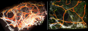
When researchers and physicians analyse tissue, for example in order to investigate any pathological changes, they often look at the tissue samples under the light microscope. However, producing meaningful images is not always easy. Researchers at the Cells-in-Motion Cluster of Excellence at the University of Münster and at the Max Planck Institute for Molecular Biomedicine in Münster have now developed a new method which, in the case of lymphoedema, can create digital 3D images of blood vessels and lymphatic vessels of entire tissue biopsies. This method will help to analyse the underlying changes of the blood and lymphatic vessels in lymphoedema in a more detailed way. “We’re doing a digital three-dimensional histopathology,” explains Dr. René Hägerling, lead author of the study which has just been published in the latest issue of the “JCI Insight” journal.

3D computer reconstruction of a healthy human skin biobsy. Spatial arrangement of blood vessels (white) and lymphatic vessels (red) is distinctly visible.
The process has involved interdisciplinary collaboration between biochemists, chemists, computer scientists, biologists and physicians. The researchers analysed three skin biopsies taken from healthy persons and one skin biopsy of a patient with lymphoedema. Using light sheet microscopy they produced thousands of individual optical sections for each sample. Using a special programming system called Voreen the researchers assembled the individual optical sections on the computer and produced a three-dimensional reconstruction of the tissue structure. The new method – called VIPAR – enables researchers for the first time to generate 3D reconstructions of skin biopsies, visualize them and extract characteristic parameters of the tissue. This method differs from traditional histological analyses, in which a tissue sample is sliced into many sections and each individual section is observed two-dimensionally.
Original publication:
Hägerling R, Drees D, Scherzinger A, Dierkes C, Martin-Almedina S, Butz S, Gordon K, Schäfers M, Hinrichs K, Ostergaard P, Vestweber D, Goerge T, Mansour S, Jiang X, Mortimer PS, Kiefer F. VIPAR, a quantitative approach to 3D histopathology applied to lymphatic malformations. JCI Insight 2017;2, DOI 10.1172/jci.insight.93424.
More information:
https://www.uni-muenster.de/Cells-in-Motion/newsviews/2017/08-28.html – The detailed story on the
Website of the Cells-in-Motion Cluster of Excellence
https://insight.jci.org/articles/view/93424 – Abstract (published in the “JCI Insight” journal)

